
|
www.efeld.com |
| The art, practice and science of Feldenkrais® Director: Robert J. Burgess BEd, PT, PhD, Feldenkrais Practitioner
|
| About The Feldenkrais Method® Rehabilitation Neuroscience Courses Products Contact |
literature review
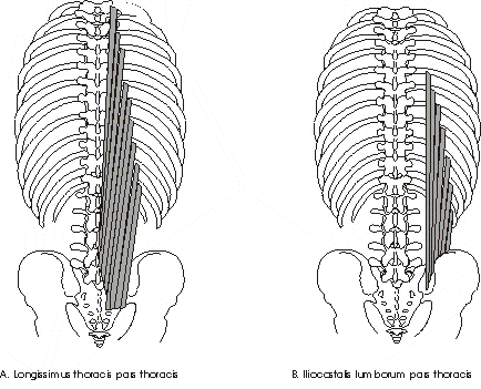
Figure 2.1 The long erector spinae muscles. The long posterior muscles of the trunk attach to the pelvis and thorax; that is they span over the lumbar spine (Macintosh and Bogduk 1987, 1991). Originating from the pelvis and some lumbar vertebrae, there is extensive insertion of fascicles to the ribs for both groups of long muscles (Figure 2.1). Sagittal and frontal standing motions of the trunk rely on these attachments for leverage and control of the thorax over the lumbar spine and pelvis. These long erector spinae muscles have been described as being suited to contributing to trunk moments and movements while the shorter lumbar muscles are better suited to control shear forces (Callaghan and McGill 1995). These significant anatomical and functional features of trunk musculature suggest that mobility and motor control of the trunk is related to the mobility and motor control of the thorax relative to the pelvis. The deep short lumbar muscles combine with the long erector spinae muscles to control trunk and hence lumbar spine motion. That one anterior trunk muscle arises from eight ribs and that one set of the posterior trunk muscles attaches to all 12 ribs suggests some role for the thorax in motor control, mobility and strength for trunk actions.
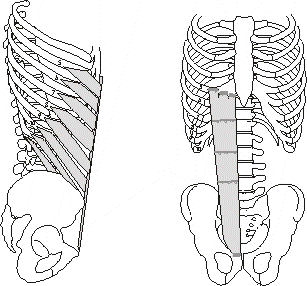
Figure 2.2 External oblique and rectus abdominus. Complete relaxation of erector spinae muscles near full flexion has been defined as the flexion relaxation response (Floyd and Silver 1950; Morris et al 1962; Kippers and Parker 1984; Ortengren and Andersson 1977; Bogduk et al 1992). Similarly, Basmajian and DeLuca (1985) also found the multifidi and rotators muscles to relax in full flexion.
|
|||||||||||||||||||||||||||||||||||||||||||||||||||||||||||||||||||||||||||||||||||||||||||||||||||||||||||||||||||||||||||||||||||||||||||||||||||||||||||||||||||||||||||||||||||||||||||||||||||||||||||||||||||||||||||||||||||||||||||||||||||||||||||||||||||||||||||||||||||||||||||||||||||||||||||||||||||||||||||||||||||||||||||||||||||||||||||||||||||||||||
|
Hindle et al 1990 |
Pearcy & Hindle 1989 |
Buchalter et al 1989 |
Dolan & Adams 1993 |
Nelson et al 1995 |
Porter & Wilkinson 1997 |
Gatton & Pearcy 1999 |
|
|
Sampling Rate Hz |
10 |
10 |
15 |
28 |
15 |
10 |
20 |
|
Sample time Seconds |
10 |
10 |
NA |
NA |
NA |
10 |
6 |
The following discussion of trunk kinematics begins with a description of the kinematics of individual trunk segments (pelvis, lumbar spine and thorax) followed by an outline of global kinematics and segmental relationships. Finally a brief review of trunk motor control is presented.
Rotation of the pelvis about the hips is a significant functional motion in human trunk action. In rising from a chair, the first motion is rotation of the pelvis and trunk about the femur (Schenkman 1990). Up to 50% of trunk flexion and extension in standing involves pelvic rotation (Mayer et al 1984; Gracovetsky et al 1995). Four to eight degrees of pelvic rotation in the sagittal plane occurs during human locomotion (Thurston and Harris 1983; Stokes, Andersson and Forssberg 1989). Sagittal plane pelvic rotation in sitting significantly effects cervical posture (Black, McClure and Polansky 1996). In the frontal and sagittal plane, pelvic rotation initiates balance control during sitting perturbations (Forssberg and Hirschfield 1994). Backward pelvic rotation initiates trunk extension from forward flexed position (Nelson, Walmsley and Stevenson 1995; McClure et al 1997). Bohannon, Gajdosik and LeVeau (1985) and Goeken and Hof (1991) reported an average of 32 and 25 degrees respectively for posterior pelvic rotation during a supine passive straight leg raising test. Pelvic rotation during trunk flexion has been shown to be altered in subjects with CLBP (Mayer et al 1984). Pelvic rotation in the sagittal plane deserves further and more complete investigation of its role in trunk kinematics in normal human function and altered function.
The term pelvic tilt has several meanings according to position, function and direction. In standing posture, pelvic tilt is a description of the static anterior-posterior position of the pelvis in the sagittal plane (Twomey and Taylor 1994). It has also been defined as the static position of the pelvis in the frontal plane (Waddell et al 1992).
Posterior pelvic rotation performed in the sagittal plane in crook lying and then held as a static position is a common reference for pelvic tilt (Kendall and McCreary 1983; Cailliet 1995). Both anterior and posterior pelvic rotation in the sagittal plane in this same position is a common position in which pelvic movements are assessed and utilized in the rehabilitation setting (Feldenkrais 1972; Kendall and McCreary 1983; Jull and Richardson 1994; Cailliet 1995). Rotation of the pelvis anteriorly and posteriorly can be performed on all fours (Jull and Richardson 1994), in standing (Cailliet 1995), or sitting (Oliver 1994) and still referred to as pelvic tilt. It can be performed in many different positions and variations for motor learning and awareness (Feldenkrais 1972, 1984).
In this study pelvic tilt was defined as anterior and posterior pelvic rotation in the sagittal plane in supine with bent legs.
Pelvic tilt in supine, though a regular rehabilitative exercise for low back pain, has not yet been the subject of a kinematic study nor is it measured clinically even though pelvic rotation has been shown to be diminished in subjects with CLBP (Mayer et al 1984). It is possible, that limited pelvic rotation in standing trunk movement may also be limited in supine. Pelvic tilt in supine is potentially a simple and easy position to observe and measure pelvic rotation in the sagittal plane. Rotating the pelvis on a firm horizontal surface provides consistent constrained conditions by which to measure pelvic tilt. Visual observation or hand held goniometric measurement of pelvic rotation in this position has the potential for a simple determination of pelvic mechanics otherwise difficult to determine in other functional positions and without sophisticated electronic equipment.
During trunk flexion, the pelvis rotates forwards about the femur during trunk flexion. Mayer et al (1984) using hand held inclinometers reported 63 degrees pelvic rotation during forward sagittal trunk motion. Similar findings have been reported by Waddell et al (1992), Esola, McClure, Fitzgerald and Siegler (1996) and Porter and Wilkinson (1997) (Table 2.2).
Table 2.2 Pelvic range of motion during sagittal plane trunk bending (in degrees). (f = female, m = male)
|
Inclino-meter |
Optoelectric device |
3Space Tracker |
||
|
Mean |
Mayer et al 1984 |
Waddell et al 1992 |
Esola et al 1996 |
Porter & Wilkinson 1997 |
|
Flexion |
63f&m |
57f&m |
70 |
58m |
|
Extension |
18f&m |
- |
- |
- |
During trunk extension the pelvis rotates backwards with the lumbar spine an average of 180 (Mayer et al 1984) (Table 2.1). Gracovetsky et al (1995) reported 4 degrees of pelvic rotation in the frontal plane accompanying 25 degrees of lumbar motion with trunk side bending.
Sagittal and frontal plane lumbar spine displacement and angular kinematics in standing are presented including ranges of motion. Using passive limits to trunk range of motion in the sagittal (experimenter applied force) and frontal planes (3 kg weight in hand) Dvorak, Panjabi, Chang, Theiler and Grob (1991a) measured displacement kinematics of the lumbar spine. For a normal or no low back pain (NLBP) population, total mean sagittal plane displacement (L1-2 to L5-S) was 54mm. In the frontal plane, displacement (L1-L2 to L4-L5) was 31.4mm (total left and right).
Flexion of the lumbar spine proceeds as an anterior vertebral rotation and an anterior translation in the sagittal plane (Kessen, During, Beeker, Goudfroois and Crowe 1984; Pearcy, Portek and Shepherd 1984). Flexion may proceed with all vertebrae moving together or a top down or bottom up or middle last sequencing (Gatton and Pearcy 1999). At the limit of flexion, the lumbar spine assumes a straight or slightly curved forwards position (Pearcy 1985) and hence the vertebral bodies become parallel to each other (Bogduk and Twomey 1991) (Figure 2.3).
Different measurement methods have produced variable data for lumbar flexion as summarized in Table 2.2. Pearcy et al (1984), using biplanar radiography, measured flexion range as 8-13 degrees for each level in 11 subjects with a total average flexion range of 51 degrees. Clinical measurements using inclinometers have determined a wide range of mean lumbar flexion from 22 to 55 degrees (Mayer et al 1984; Mellin 1990). Goniometric recordings for lumbar flexion from Mayer et al 1984 were between the reported electro-magnetic device and radiographic results whilst Mellin's (1990) goniometric recordings results seem extremely low. Mellin (1990) measured 52 females and males with a mean age 21.4 ± 1.6 years while Mayer's subjects were aged 19-51 with a mean age of 31 years. Hence age and subject were unlikely to affect these large differences in lumbar flexion range. Perhaps skill of inclinometer use may be a factor (Portek, Pearcy, Reader and Mowat 1983). Electronic goniometric measurement produces spinal ranges of motion very similar and sometimes a little larger than radiographic results (Pearcy 1985; Buchalter, Parpianpour, Viola, Nordin and Kahanovitz 1988; Pearcy and Hindle 1989; Nelson et al 1995).
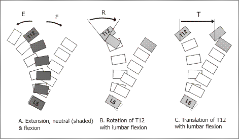
Figure 2.3 Lumbar spine kinematics Schematic representation of the lumbar spine kinematics in the sagittal plane. A. Lumbar extension, E (to left), neutral stance (shaded) and flexion, F (to right). B. Anterior vertebral rotation of T12 during lumbar flexion. C. Anterior vertebral translation of T12 during lumbar flexion. Lumbar range of translation and rotation during trunk bending is obtained by subtracting the translation and rotation for T12 from the translation and rotation for the pelvis (Figure adapted from Pearcy 1985).
Table 2.2 Lumbar flexion range of motion (in degrees)
|
3Space Isotrak |
Biplanar radio-graphy |
Inclino-meter |
||||||
|
Hindle et al 1990 |
Pearcy & Hindle 1989 |
Buchalter et al 1988 |
Pearcy 1985 |
Dvorak et al 1991a |
Mayer et al 1984 |
Mellin 1990 |
||
|
Mean Flexion |
67 f 74m |
76 |
56 |
51 |
80 |
55 |
22 f 27 m |
|
Extension of the lumbar spine is not as frequently performed in daily life activities as flexion and therefore perhaps has not been the subject of as much research (Crenna et al 1987). Extension has been stated to occur as the converse of flexion (Bogduk and Twomey 1991; McClure et al 1997) ie with posterior vertebral rotation and posterior translation.
McClure et al (1997) investigated extension of the lumbar spine from a flexed position and described the return to upright stance from full flexion as the reverse of flexion, however Nelson et al (1995) found pelvic rotation to initiate trunk extension while the lumbar spine actually flexed initially.
The range of extension has been measured as consistently less than flexion (Mayer et al 1984; Pearcy et al 1984; Pearcy 1985; Buchalter et al 1989; Pearcy and Hindle 1989; Hindle, Pearcy, Cross and Miller 1990). Only Mellin (1990) recorded extension as nearly twice that of flexion of other studies (Table 2.3) which may perhaps be explained by the large variability found when using inclinometers (Portek et al 1983).
Table 2.3 Lumbar extension range of motion (in degrees)
|
3Space Isotrak |
Biplanar radio-graphy |
Inclino-meter |
||||
|
Buchalter et al 1988 N=60 33f 27m |
Pearcy & Hindle 1989 N=10m |
Hindle et al 1990 N=80 40f 40m |
Pearcy 1985 |
Mayer et al 1984 N=13 6f 7m |
Mellin 1990 N=103 48f 55m |
|
|
Mean Flexion |
22 |
23 |
24f 21m |
16 |
27 |
40f 44m |
Taylor and Twomey (1980) measured large numbers of 437 living subjects using a spondylometer. They did not differentiate between flexion and extension, instead reporting a mean combined sagittal rotation score of 42 degrees while Dvorak et al (1991a) measured a mean 80 degrees total sagittal plane motion using radiographic techniques. Dvorak et al (1991a) measured a passive (experimenter force at end of range) limit to flexion and extension. Buchalter et al (1989), Pearcy and Hindle (1989) and Hindle et al (1990) reported 78 degrees, 99 degrees and 95 degrees respectively for total active trunk sagittal plane bending. Hence the radiographic measurements of Dvorak et al (1991a) and the electromagnetic devices are comparable, however, Taylor and Twomey's (1980) lumbar sagittal motion is extremely low by comparison. This difference illustrates the errors inherent in the inclinometer method of spinal motion measurement (Portek et al 1983).
With lateral flexion of the lumbar spine, the upper vertebra rotates and translates in the frontal plane in the direction of motion (White and Panjabi 1978). As with all other lumbar motions, lateral flexion also eludes accurate determination by clinical measuring devices and can only be said to range between 35 and 60 degrees (Table 2.4). Radiographic techniques again result in low values for lumbar motion (Pearcy and Tibrewal 1984), while clinical measures vary from 42 degrees using an inclinometer (Mellin 1990) to 62 degrees utilizing the 3Space Isotrak (Hindle et al 1990) and automated video (Vachalathiti, Crosbie and Smith 1995). Though some accompanying lumbar rotation occurs with lumbar lateral flexion (Hindle et al 1990), the primary motion only will be investigated in this study.
Table 2.4 Lumbar spine frontal plane range of motion (in degrees)
|
Radio-Graphy |
3Space Isotrak |
Inclino-meter |
Auto-mated video |
||||
|
Authors |
Pearcy & Tibrewal 1984 |
Dvorak et al 1991a |
Hindle et al 1990 |
Pearcy & Hindle 1989 |
Buchalter et al 1988 |
Mellin 1990 |
Vachalathiti et al 1995 |
|
N=10 10m |
N=41 18f 23m |
N=80 40f 40m |
N=10 10m |
N=60 33f 27m |
N=103 48f 55m |
N=100 54f 46m |
|
|
Mean Lateral Flexion |
35m |
58 |
62f 58m |
56m |
47 |
42f 45 m |
62 |
The thorax as the composite of 24 ribs, a sternum and 12 thoracic vertebrae can be considered as a separate segment of skeletal motion with a range of movement (White 1969). Over 70 articulations and 204 possible combinations for movement ensure some mobility in the thorax (Anderson 1982). Based on experimental evidence, mathematical models and clinical experience, Lee (1993) proposed a biomechanical model for the thorax illustrated in Figure 2.3. This model is utilised here to describe sagittal and frontal plane displacement and angular kinematics of the thorax.
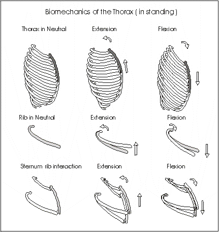
Figure 2.4 Biomechanical model for the thorax. Sagittal plane thorax rotation. A. Neutral position of the ribs and sternum in standing. B. Extension of the thorax is accompanied by a posterior rotation and elevation of the ribs and presumably the sternum. C. Flexion proceeds with anterior rib rotation and sternal depression (adapted from Lee 1993).
Occurring with a sagittal rotation and translation motion of thoracic vertebrae (Panjabi and White 1978), thorax flexion and extension proceeds with accompanying rib movements (Lee 1993).
As shown in Figure 2.4, theoretical extension of the thorax is coupled with a posterior rotation of the ribs (Lee 1993). Concomitantly, the sternum would follow this action and elevate and rotate backwards with extension. Conversely, with flexion, the ribs rotate anteriorly producing a depression of the sternum. This theoretical model for thorax motion is presumably valid in both supine and standing motion in the sagittal plane.
Table 2.5 Thorax sagittal and frontal plane range of motion (in degrees) * summated values from authors results. (f = females, m = males).
|
Device |
Literature Review |
Inclino-meter |
3Space Isotrak |
||
|
Study |
White 1969 |
White & Panjabi 1978 |
O'Gorman & Jull 1987 (in sitting) |
Mellin 1990 |
Buchalter et al 1989 |
|
Sample size |
N=120w |
N=103 48w 55m |
N=60 33w 27m |
||
|
20.4* |
- |
22 |
60m 60f |
20 |
|
|
Extension |
13.6* |
- |
20 |
4m -2f |
20 |
|
Combined flexion-extension |
34 |
76 |
42* |
58-60* |
40 |
|
Lateral flexion |
- |
72* |
25f |
70m 64f |
40f&m |
Total thorax sagittal range of motion has been recorded in vitro as 34 degrees (White 1969) with a flexion to extension ratio of 3:2. Panjabi and White (1978), however, from a literature review quote a total 76 degrees for sagittal thorax motion. They reported 4 degrees in each of the upper inter-vertebral levels, 6 degrees in the middle levels and 12 degrees in each of the lower levels (Table 2.5). In contrast, Buchalter et al (1989) found a total sagittal range of motion of 40 degrees equally distributed between flexion and extension. Mellin (1990) reported 60 degrees for flexion and only minimal extension of 4 to -2 degrees.
Motion of the thorax in the frontal plane results in frontal rotation of the thoracic vertebra and is theoretically accompanied by approximation of rib margins on the same side and rib separation on the opposite side. It is possible that the approximation of rib margins limits the motion of lateral flexion prior to vertebral limitations (Lee 1993). White and Panjabi (1978) quote the range of motion of lateral flexion to be fairly consistent throughout the length of this spinal segment at a mean of 6 degrees, though increasing slightly at lower levels. Buchalter et al (1989) found an overall average of 40 degrees in a group of 20 to 50 year olds. In sitting, O'Gorman and Jull (1987) reported 37 degrees lateral flexion in the 22 to 29 age group, while Mellin (1990) recorded 70 degrees for males and 64 degrees for females.
During lateral flexion, the thoracic vertebral spinous processes also rotate in the horizontal plane towards the concavity of the curve (Gregersen and Lucas 1967; White 1969). While lateral flexion may be accompanied by rotation, the primary movement of rotation in the frontal plane is much greater and will be considered in this study without its coupled motion.
Lumbar, thoracic and pelvic motion is most often measured individually and few studies have investigated the motion of these segments simultaneously. Presented below are some studies that have measured trunk segment motion simultaneously. These studies include measurement of pelvic and lumbar motion during trunk flexion (lumbopelvic rhythm or lumbopelvic ratio) and extension and thorax motion recorded simultaneously during trunk motion.
The ratio of lumbar to pelvic contributions to trunk flexion has been investigated (lumbar range / pelvic range = L-P ratio). Mayer et al (1984) reported lumbopelvic ratio (L-P ratio) over two periods of trunk flexion: up to 90 degrees flexion and 90-120 degrees and found L-P ratios of 1.72 and 0.17 respectively (Table 2.6). Lumbar movement was greater than pelvic motion during the first 90 degrees of trunk flexion and then pelvic motion predominated in the later ranges of trunk flexion. These results have been repeated in other studies using more sophisticated electronic equipment to find similar L-P ratios (Esola et al 1996; Porter and Wilkinson 1997; Granata and Sanford 2000) (Table 2.6). Authors vary in what reference point to use for the lumbar spine. The upper limit for the lumbar spine has been selected as L1, T12-L1, T12, T10 and C7 which may produce varying results depending on thorax contributions to trunk bending (Table 2.6). Presumably, in choosing T10 or C7 for the upper limit for the lumbar spine authors are making the assumption that the thorax does not contribute to trunk bending. This may not be the case. Gracovetsky et al (1995) reported a continuous constant equal L-P ratio throughout trunk flexion and actually calculated a trunk-pelvic ratio rather than an L-P ratio (Table 2.6). Similarly, Granata and Sanford (2000) in determining L-P ratio defined the lumbar spine as L5 to T10. This may be different to the actual true L-P ratio. Nelson et al (1995) reported that at 50% of full trunk flexion, the lumbar spine had flexed to 94% of its maximum and the pelvis had reached 84% of its maximum. These figures are difficult to compare to other studies using direct ratios between lumbar and pelvic motion.
Farfan (1975) suggested that pelvic and lumbar movement occurred sequentially while Nelson et al (1995) reported lumbo-pelvic rhythm to be more sequential during lifting (extension) and more simultaneous during lowering (flexion) a 9.5kg box to 90% to full trunk flexion. Pelvic backward rotation always preceded lumbar extension during lifting a 9.5kg load (Nelson et al 1995). Granata and Sanford (2000) found simultaneous motion for these segments during trunk bending.
Load may alter lumbo-pelvic mechanics. Granata and Sanford (2000) reported the lumbar spine to contribute 70 percent of forward bending (pelvic 30%) which increased when weight was added to the task. This contrasts with Gracovetsky et al (1995) who found no affect on spinal coordination with loading.
Few studies have reported simultaneous pelvic, lumbar and thoracic motion. One spinal motion study did actually record cervical, thoracic and lumbar motion simultaneously for standing flexion, extension, rotation and lateral flexion using an electromagnetic sensing device (Buchalter et al 1989). All spinal segments, cervical, thoracic and lumbar contributed to each trunk movement. Interestingly, only for flexion was lumbar range of motion considerably more than thoracic motion. For extension and lateral flexion, thoracic and lumbar motion was almost equal, and for rotation, thorax motion was more than four times that of lumbar motion (Buchalter et al 1989).
Tully and Stillman (1997) using computer aided video analysis studied two groups of subjects; one group that could touch their toes and one that could not. Lumbar and thorax movements were measured during a toe touching test. The authors determined five different patterns of thorax movement during trunk flexion. (Dividing the motion into three stages, initial, middle and final thirds of the test, patterns were defined as follows: flexion-extension-extension, flexion-extension-flexion, extension-extension-flexion, flexion-flexion-flexion and extension-flexion-flexion). The lumbar spine consistently flexed during the test. Interestingly, subjects who could successfully reach their toes utilized more thorax extension (mean 4.0 ± 14.7 degrees) whilst the unsuccessful toe touchers used more thorax flexion (mean = 4.0 ± 10.2 degrees and overall mean for both groups = 0.0 ± 12.9 degrees).
Table 2.6 Lumbo-pelvic ratio *Lumbar-pelvic ratio = lumbar motion/pelvic motion.
|
Measurement device |
Inclinometer |
Opto-electric mdevice |
Tracker |
Electro-magnetic device |
||
|
Study |
Mayer et al 1984 |
Gracovetsky 1995 |
Esola et al 1996 |
Porter & Wilkinson 1997 |
Granata & Sanford 2000 |
|
|
Upper limit for lumbar spine |
T12-L1 |
C7 |
T12-L1 |
L1 |
T10 |
|
|
Range of trunk flexion |
Lumbo-pelvic ratio* |
|||||
|
0-30 |
- |
1 |
2 |
- |
2.4 |
|
|
30-60 |
- |
1 |
1 |
- |
2.4 |
|
|
60-90 |
- |
1 |
0.5 |
- |
1.8 |
|
|
50% flexion |
- |
1 |
- |
- |
- |
|
|
Full flexion |
- |
1 |
- |
- |
- |
|
|
0-90 |
1.7 |
1 |
- |
2 |
- |
|
|
90-120 |
0.17 |
1 |
- |
0.33 |
- |
|
It is difficult to determine adequate reasons for these discrepancies in flexion motion. Methods, pitfalls and developments in measuring spinal motion are discussed in section 2.5 Measurement of Spinal Motion. It is also possible that different tasks demand different contributions of thorax motion.
Cavanaugh, Shinberg, Ray, Shipp, Kuchibhatla and Schenkman (1999) investigated trunk kinematics during an asymmetrical functional reach task. Subjects were required to reach as far forward as possible without falling. Young subjects (N = 34, mean age = 28) reached significantly further than the elderly subjects (N = 32, mean age = 70 years). The elderly displayed significantly less thorax rotation and centre of mass (COM) displacement.
Many authors have previously reported the thorax to contribute little to spinal movement (Davis et al 1965; Munro 1965; Grieve 1988; Cailliet 1995; Lindh 1989) and it is usually modelled as a rigid segment (Gracovetsky and Farfan 1986). This assumption of thorax rigidity may be justifiable when considering the minimal motion found by studies like Tully and Stillman (1997) but not when considering the 20-60 degrees reported by Buchalter et al (1989) and Mellin (1990). What seems possible is that under some circumstances of trunk movement the thorax is required to move minimally and under others it moves considerably more depending on the task and method of performing that task. This was observed by Cavanaugh et al (1999) during the previous described functional reach task. Pre-test trunk range of motion measurements were taken for the thoracic and lumbar spines. During the maximal reach task, only 30 and 50 percent of available rotation and lateral flexion respectively were utilised.
Recently, reports on spinal modelling and stability have acknowledged the role of the long erector spinae muscles attaching along the length of the rib cage (Bergmark 1989; McGill 1992; Kiefer, Shirazi-Adl and Parnianpour 1998). It seems possible that if long muscles of the trunk attach from the pelvis to each rib that some leverage in muscle action may be gained from a flexible rib cage. However in these studies the thorax is modelled as a rigid segment and the question of thoracic mobility and leverage was not explored. One possible reason for modelling the thorax as a rigid segment is to reduce the complexity of the analysis in favour of a simplistic workable mathematical model.
Jull and Richardson (1994) comment that with a posterior pelvic tilt, the anterior rib cage depressed downwards. Abdominal muscles contracting to tilt the pelvis, may affect the position of the sternum and ribs pulling them downwards. Similarly, motion of the pelvis may be transmitted through the lumbar and thoracic vertebrae to determine sternal and thorax kinematics (Figure 2.5).
Activation of the long erector spinae muscles to produce anterior pelvic tilt and hence extension of the lumbar spine may also extend the thorax and tilt the sternum upwards. Black et al (1996) reported a relationship in sitting between pelvic motion and resultant cervical spine posture but did not determine the intermediary thorax kinematics.
Feldenkrais (1972) described a corresponding matched motion of the pelvis, thorax and head with pelvic anterior and posterior tilting. Feldenkrais proposed that coordination between the head and pelvis via the length of the spine and chest is essential for efficient strain free spinal movement. Simultaneous supine pelvic rotation and corresponding thorax motion has not been previously investigated.
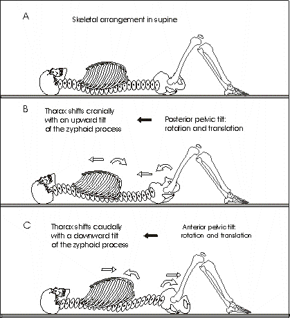
Figure 2.5 Pelvic tilt and the thorax. Schematic representation of pelvic tilt. Posterior and anterior pelvic tilt produces both displacement and angular kinematics. A. Posterior pelvic tilt pushes through the vertebrae to produce a cranial translation and a depressive rotation of the sternum. B. Anterior pelvic tilt pulls on the vertebrae to produce a caudal translation and a cranial rotation of the sternum.
Lifting a box from several different platforms (floor, knee, hip and chest height) has been a frequent method of studying the biomechanics of the spine for nearly 50 years (Floyd and Silver 1955; Nussbaum et al 2000). Whilst trunk kinematics during lifting has been studied extensively, investigation of trunk segmental interactions is sparse. For example, Tsuang, Schipplein, Trafimow and Andersson (1992) modelled body segments during a lifting task. Markers on the hand, elbow, greater trochanter, humerus, knee and ankle provided data for a five bar linkage model. However, no segmental trunk information was allowed for in this methodology. Similarly, Khalaf et al (1999) developed a methodology for evaluating lift characteristics, using markers on the ankle, knee, hip, shoulder and wrist. A model was formulated from these markers and hence again the trunk was modelled as one rigid segment.
Nussbaum et al (2000) measured torso kinematics during a lifting task using markers on L5/S1, T10 and T4. The torso was defined as T4 and T10 relative to the pelvis; however only one score for torso kinematics was presented without identifying what segment was considered to be the torso.
Interactions between voluntary and postural kinematics during perturbed lifting has been investigated in an asymptomatic population (Oddsson, Persson, Cresswell and Thorstensson 1999). Subjects were required to lift a 20kg box from floor to desk and were perturbed in the anterior and posterior directions. The study found large erratic erector spinae muscles activity during a backward perturbation. Oddsson et al (1999) proposed that this rapid switch from voluntary contractions to postural reactions as a possible mechanism of injury to the low back. Again, inter-segmental trunk kinematics was not determined in this study.
Gracovetsky, Kary, Pitchen, Levy and Ben Said (1989) found increased EMG activity in the spinal muscles and increased compressive stress within the spine, when the lumbosacral lordosis of the spine increased or decreased beyond an optimal level during a lifting task. The model used predicted that for every angle of flexion, there was a unique amount of pelvic tilt to minimize compressive forces within the lumbar spine.
Several studies of trunk kinematics during lifting tasks have measured lumbar spine motion as being between T10 and the pelvis and sometimes the trunk as the motion from the pelvis to T10 (Marras, Lavender, Leurgans, Rajulu, Allread, Fathallah and Ferguson 1993; Fathallah et al 1998; Granata and Sanford 2000). In these studies the lumbar spine is modelled with three extra vertebrae and the trunk is missing 10 thoracic vertebrae. If all studies consistently use this method then perhaps this protocol is a valid one. Many other studies, however, have used either T12 (Buchalter et al 1989; Waddell et al 1992; Porter and Wilkinson 1997) or L1 (Pearcy and Hindle 1989; Hindle et al 1990; Dolan and Adams 1998) or both (ie between T12 and L1) (Mayer et al 1984; Nelson et al 1995; Esola et al 1996) to represent the upper limit of the lumbar spine. In lifting and balance studies, the trunk is most often defined as being between C7 or the shoulder and S2 or the greater trochanter (Anderson 1982; Allum, Bloem, Carpenter, Hullinger and Hadders-Algra 1998; Diener et al 1990; Alexander et al 1992; Tsuang et al 1992; Khalaf et al 1999; Oddsson et al 1999; Rietdyk, Patla, Winter, Ishac and Little 1999). Defining trunk segments and assumptions about movement contributions of the thorax could influence results. To save confusion, inconsistent or incorrect results, it may be better to maintain a consistent anatomically correct definition for kinematic studies of lumbar and trunk motion. The current study defined the trunk as that segment between C7 and the pelvis and the lumbar spine as between the pelvis (S2) and T12 (Study 1) or L1 (Study 2).
Trunk action from standing involves a coordinated sequencing of most of the body segments: the head, trunk, pelvis and legs (Crenna et al 1987; Pedotti et al 1989; Oddsson 1990). Babinski (1899) first reported the accompaniment of hip and knee motion with trunk bending. In the sagittal plane trunk bending proceeds as a coordinated action of the head and trunk in the direction of the intended movement and the pelvis and knees in the opposite direction (Figure 2.6). The displacement of the pelvis and legs opposite to the intended movement of the trunk occurs to maintain postural equilibrium (Crenna et al 1987). With voluntary action in standing, muscle activation and body segment kinematics occur to achieve the intended command but also to control posture and balance. Similarly, Belenki, Gurfinkel and Palistev (1967) showed that raising an arm was preceded by a backward movement of the trunk and legs aimed at minimizing the disturbance of balance due to the movement. The accompaniment of trunk bending with these adjustments in posture is called anticipatory postural adjustments (Massion 1992). The intended movement may also cause reaction forces and inter-segmental interactions through the posturo-kinematic chain resulting in associated movement separate from the voluntary intention (Massion, Alexandrov and Vernazza 1998).
It has been shown that a burst of muscle activity in the lower leg, thigh and trunk precedes the onset of movement of that segment (Crenna et al 1989; Oddsson and Thorstensson 1986). Hence, because muscles act before movement rather than in response to movement, it is proposed that trunk axial synergies are centrally controlled and of the anticipatory postural adjustment type (Massion et al 1998).
Using a rigid three link model Ramos and Stark (1990) determined that the backward movement of the pelvis and legs during the forward motion of the trunk occurred as a necessary result of the mechanical relationships between these structures as well as the result of anticipatory postural adjustments for maintenance of postural stability.
The relationship between ankle, knee and hip angle has been shown to be linked by fixed ratios that could be reduced to one controlling parameter (Alexandrov, Frolov and Massion 1998). These authors suggested that only one degree of freedom was needed to control trunk bending and proposed two hypotheses for explaining this occurrence. This fixed ratio could result from passive biomechanical constraints or a central command adjusted to the biomechanical properties of the individual. That is to say that both neurological and mechanical factors may determine trunk kinematics.
Lumbar lordosis and trunk angle have been determined as strictly correlated suggesting that the CNS controls the degree of freedom of the spine (Mitnitski, Yahia, Newman, Gracovetsky and Feldman 1998). Mitniski et al (1998) measured lumbar and trunk angle while subjects lifted varying weights (bar 11-68kg) from 10cm height to mid thigh to find that lumbar lordosis and trunk angle were highly correlated and therefore limiting the degrees of freedom to be controlled by the CNS. Hence the results of Mitniski et al (1998), Alexandrov et al (1998) and Ramos and Stark (1990) suggests that CNS must control both trunk angular and displacement kinematics.
Trunk motion follows a bell shaped velocity curve (Oddsson 1988). During slow sagittal (one second for forward flexion) and frontal plane movements of the trunk, motion proceeds sequentially craniocaudally and during fast motions the sequencing is simultaneous (Thorstensson et al 1985; Crenna et al 1987). Hence speed of motion may affect motor control of the trunk.
During trunk flexion and lateral flexion, pelvic action, lumbar elongation and lordosis proceed in consistent patterns despite loading and range of movement (Gracovetsky et al 1995). Similarly, Vernazza, Alexandrov and Massion (1996) showed that the final position of the centre of mass (COM) during a trunk bending task remained constant despite changes in load applied to the shoulders. Hence subjects predicted the effect of additional load and adjusted the kinematics accordingly to maintain the COM within the support base.
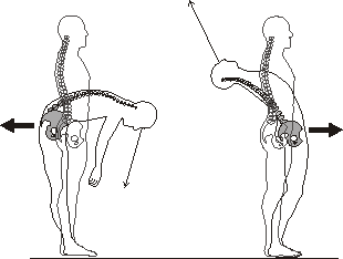
Figure 2.6 Coordination of the trunk and pelvis during axial bending. With forward flexion, the pelvis and knees displace backwards, while the trunk and head translate forwards. While with extension, the pelvis and knees translate forwards and the head and trunk displace backwards. The eyes lead trunk motion.
Trunk motion can be performed consistently in similarly reproducible patterns. Six repeated trials with the same subject produced consistent motion patterns for the head, trunk, pelvis and legs during trunk extension (Oddsson 1988). Even over two days trunk bending in the sagittal plane has shown to be consistent as illustrated by coefficient of variance scores less than 0.20 for pelvic and lumbar in 10 subjects (Nelson et al 1995). These features of trunk motion suggest a well defined pattern.
The position of C7 relative to the pelvis in the sagittal and frontal planes in standing and lying down is well controlled by the nervous system. Standing and supine positioning of C7 relative to the pelvis in the frontal plane is highly consistently controlled (Jakobs, Miller and Schultz 1985).This study found standing subjects were able to return C7 to its neutral upright position to within 3.1mm or 0.3 degrees subtended at the sacrum after passive and active repositioning in standing and sitting in the frontal plane. In the horizontal plane subjects could rotate the trunk by rotating the shoulders relative to the hips or the hips relative to the shoulders to 2.8 ± 1.2 degrees and 2.9 ± 1.4 degrees respectively. Through each stride in human gait, the trunk remains vertical to within ±1.5 degrees in the sagittal and frontal planes (Winter and Eng 1995).
Trunk motion is known to be altered in some movement trained individuals (Pedotti et al 1989; Mouchnino, Aurenty, Massion and Pedotti 1993). Dancers maintain trunk alignment to the vertical during standing leg abduction to 45 degrees while untrained individuals tilt their trunk significantly with abduction of the leg (Mouchnino 1993). Gymnasts are able to perform trunk movements on a narrow beam without loss of balance while untrained individuals cannot (Pedotti et al 1989). Muscle synergies during backward trunk bending and control of COM in trained subjects showed more highly flexible and adaptable patterns than the untrained.
Generally, range of spinal motion decreases significantly with age (Taylor and Twomey 1980; Twomey and Taylor 1983; O'Gorman and Jull 1987; Hindle et al 1990; Gracovetsky et al 1995; Vachalathiti et al 1995) (Tables 2.7 and 2.8). There have been no reported gender differences for thorax motion (Buchalter et al 1989) while lumbar spine studies have reported less lumbar flexion in females (Wolf, Basmajian, Russe and Kutner 1979; Burton and Tilloston 1988; Hindle et al 1990; Vachalathiti et al 1995).
With advancing age, the pelvic contribution to forward bending increases, corresponding to a decrease in lumbar motion and a decrease in the ability to reduce lumbar lordosis. There is little change in pelvic motion over the fourth and fifth decades (Gracovetsky et al 1995).
O'Gorman and Jull (1987) investigated the thoracic motion of 120 adult females in sitting. As illustrated in Table 2.7, flexion decreased significantly from the twenties to the thirties, remained consistent over the next two decades (30-50 years) and then dropped significantly beyond age 50. Extension decreased significantly from the third through the fifth decade but then altered minimally beyond the fifth decade. Buchalter et al (1989) found no significant relationship between age, gender and range over the third and fourth decade for thorax motion.
Table 2.7 Thorax mobility with age in females. (L = left, R = right) (Age in years, range in degrees, O'Gorman and Jull 1987).
|
Age (N= 120) |
||||
|
years decade |
22-29 3rd |
30-39 4th |
40-49 5th |
50-59 6th |
|
Flexion |
33 |
25 |
23 |
16 |
|
Extension |
37 |
29 |
21 |
20 |
|
Lateral flexion |
37 |
L 30 R 33 |
26 |
22 |
Hindle et al (1990) investigated lumbar mobility and found a consistent loss of range of lateral flexion with age in both sexes, while extension was decreased in females only (Table 2.8). Flexion did not significantly reduce with age in either gender group. Vachalathiti et al (1995) studied three age groups (20 to 25, 36 to 59 and >60) and as illustrated in Table 2.8, flexion and lateral flexion did not significantly change with age until beyond 60 years. Again males had more flexion than females which has also been previously reported (Wolf et al 1979; Burton and Tilloston 1988; Hindle et al 1990).
Table 2.8 Lumbar mobility with age and gender *significance level p = 0.05, ** p = 0.01. significant for > 60 compared to < 60 years significant for > 60 compared to >35 years.
|
study |
Hindle et al 1990 |
|||||
|
females |
males |
|||||
|
age range of motion |
20-29 |
30-39 |
40-49 |
20-29 |
30-39 |
40-49 |
|
flexion |
59** |
70 |
64* |
75 |
73 |
77 |
|
extension |
32 |
24 |
20 |
26 |
17 |
24 |
|
lateral flexion |
62 |
54 |
53* |
58 |
53 |
47 |
|
study |
Vachalathiti et al 1995 |
|||||
|
females |
males |
|||||
|
age range of motion |
20-35 |
36-59 |
>60 |
20-35 |
36-59 |
>60 |
|
flexion |
39 |
37 |
32 |
48 |
46 |
33 |
|
lateral flexion |
66 |
62 |
49 |
67 |
62 |
53 |
The problem of persistent CLBP is so great that it eludes a clear pathophysiological definition and is currently only recognizable by its persistence despite investigation and treatment. For example, CLBP has been defined as back pain persisting for longer than three months (Waddell et al 1992), pain persisting for greater than 49 days (Quebec-Task-Force-on-Spinal-Disorders 1987) or two years work loss (Mellin 1988).
CLBP is also described as the end stage of spine problems (Mayer 1991) and in terms of disability and societal costs (Frymoyer and Cats-Baril 1991; Tulder et al 1995). It is most prevalent and persistent through the 4th and 5th decade of life (30 to 50 years) (Long, BenDebba and Torgenson 1996). A person with CLBP and off work for six months has a 50% chance of a return to work, while only a 10% chance after 12 months and no chance after two years (Beals and Hickman 1972). Hence once low back pain becomes established it becomes more difficult to ameliorate.
CLBP is a major issue for the individual, health professions and the community. Ongoing determination of contributing factors is necessary to better define and manage the problem. Therefore it is imperative that a better understanding of the contributing factors be developed. One development in the understanding of CLBP has been the identification of an altered kinematic relationship between pelvic and lumbar forward bending in subjects with CLBP (Mayer et al 1984; Dolan and Adams 1993; McClure et al 1997; Porter and Wilkinson 1997). It is possible that other contributory kinematic relationships exist between motion segments adjacent to the lumbar spine.
The skeletal segment immediately adjacent superiorly to the lumbar spine is the thorax. As described earlier, the powerful muscles of the trunk span over the lumbar spine from the pelvis to the thorax and hence all three segments coordinate in trunk function and may each contribute to altered spinal kinematics relationships in CLBP.
There are many clinical tests that successfully differentiate the low back pain sufferer from normal individuals, however as yet, none can differentiate low back pain from CLBP (Waddell, Allan and Newton 1991). The most common positive clinical tests for CLBP are reduced flexion, extension (pelvic plus lumbar rotation), lateral flexion, straight leg raising range of motion and sit up weakness (Waddell et al 1992). Hip joint immobility (Mellin 1988; Dolan and Adams 1993) and an altered lumbopelvic rhythm (Mayer et al 1984; Porter and Wilkinson 1997) have also been identified in CLBP sufferers.
Those physical parameters that differentiate low back pain from CLBP have not yet been defined. Possibly there may not be any physical characteristics that indicate that back pain will become chronic, persistent and unresponsive to treatment. Other parameters such as social and environmental features may be more involved in chronicity than physical characteristics.
One of the problems in identifying significant objective clinical measures is the difficulty in differentiating between physical disability and pain inhibition, fear avoidance, psychological distress and illness behaviour (Waddell et al 1992). These features are difficult to control for and may influence results of any study. Avoiding the selection of CLBP sufferers currently experiencing an acute bout of pain will possibly lessen pain inhibition to some degree, reduce fear avoidance and limit psychological distress factors. Otherwise these features remain limiting factors.
Many individuals with CLBP have psycho-social issues (Waddell 1992; Keefe, Beckham and Fillingim 1991; Feuerstein and Beattie 1995). CLBP is not just a medical condition nor is it just a psychological problem. Loeser (1991) maintained that interpersonal and environmental factors leading to unresolved stress are the major determinants of disability associated with pain. These issues are beyond the scope of this study and will not be considered further.
Aside from the obvious complexity of CLBP, the majority of sufferers have a mechanical component (Bogduk and Twomey 1991) with measurable physical parameters (Waddell et al 1991). Range of motion remains a significant parameter for assessment of back pain in the clinic and the laboratory (Helliwell et al 1992; Mayer 1991; Waddell et al 1991).
Though pelvic rotation during trunk motion has been shown to be diminished in subjects with CLBP (Mayer et al 1984; McClure et al 1997; Porter and Wilkinson 1997) there exists no clinical measurement tool to determine this dysfunction, and though pelvic tilt is reported as critical to lumbar stability (Kendall and McCreary 1983; Jull and Janda 1987; Cailliet 1995; Jull and Richardson 1994) no studies have actually measured pelvic rotation in supine in either an asymptomatic or CLBP population.
Dvorak et al (1991b) found significantly less lumbar displacement with trunk flexion for a low back pain population (N= 27, mean age 40, 46mm) compared to a normal population (54mm).
Several studies have found lumbar spine flexion to be reduced in subjects with low back pain (Mayer et al 1984; Pearcy et al 1985; Burton, Tillotson and Troup 1989; Dvorak, Panjabi, Novotny, Chang and Grob 1991b). Flexion has been more extensively studied than extension (Mayer et al 1984; Pearcy et al 1985).
In contrast, other studies have found no significant difference between lumbar flexion in pain free subjects and subjects with CLBP (Rae, Venner and Waddell 1981; Waddell et al 1992). Waddell et al (1992) using inclinometers did however find pelvic flexion (forward pelvic rotation with trunk flexion) and total flexion (pelvic and lumbar spine) to be significantly reduced in subjects with CLBP. Similarly, Dolan and Adams (1993) and Porter and Wilkinson (1997) found pelvic flexion to be significantly reduced in subjects with CLBP. Hence forward flexion was affected by back pain but this may not be expressed in the lumbar spine.
Studies of elite athletes have not demonstrated any significant loss of lumbar motion with CLBP (Klein, Snyder-Mackler, Roy and DeLuca 1991; Sward, Eriksson and Peterson 1990). Dvorak et al (1991b) investigated three groups of subjects: those with low back pain, those without and athletes. The group with low back had significantly less sagittal angular and displacement range of motion (combined flexion and extension) than the normal group, while athletes had significantly greater motion than normals. Athletes tend to maintain flexibility of leg and trunk muscles and hence perhaps this reduces any measurable loss of range related to back pain. Similarly, demand on the athlete's body exceeds normal function and therefore perhaps minor differences in range become significant in function but not to measurable devices.
Mellin (1990) found lumbar extension and lateral flexion to be significantly reduced in males with CLBP but not in females. Historically, lateral flexion is included as a significant positive test for low back pain (Weitz 1981; Waddell et al 1992) and has been found to be sometimes significantly reduced in subjects with low back pain (Troup, Martin and Lloyd 1981; Mellin 1990; Waddell et al 1992).
L-P ratio has been reported as increased in subjects with CLBP, that is, lumbar motion during trunk flexion is increased relative to pelvic motion. Mayer et al (1984) found mean pelvic flexion to decrease from 66 degrees in a normal group to 42 degrees in a CLBP population. Dolan and Adams (1993) found pelvic motion to be reduced more than lumbar motion. Pelvic motion decreased by 20 degrees compared to seven degrees for lumbar flexion. Porter and Wilkinson (1997) found 70 percent of a CLBP population to have reduced lumbar but not pelvic motion and 30 percent to have diminished pelvic but not lumbar motion.
Hip joint range of motion is the result of femur movement relative to the pelvis. Alternatively, the femur can be held fixed while the pelvis is moved relative to the femur. Performed in the sagittal plane this would result in posterior and anterior pelvic tilting. It is possible that any hip restriction observed in the sagittal plane may also be reflected by some change to pelvic motion in the same plane. Hence lumbar spine and hip joint restrictions may affect pelvic tilt.
Mellin (1986, 1988) investigated the mobility of the hip joints in subjects with CLBP. Reporting reduced hip flexion, extension and internal rotation in subjects with low back pain, Mellin (1988) listed several possible factors contributing to this relationship:
1) a decrease in general activity associated with low back pain which may cause limitations in hip mobility
2) spasm from back pain and pathology via reflexes may cause restricted movement patterns in the hips
3) hip restriction may increase loads on the lumbar spine
4) hip stiffness may be aetiologically associated with low back pain.
Mellin (1987), using Schrober's tape method, found thoracolumbar (lumbar plus thorax) to be more strongly correlated with CLBP than the lumbar spine alone. This suggests that CLBP motion characteristics are global as well as local and possibly related to thorax mechanics. Mellin (1990) recorded thoracic spinal motion in a study of young adults with CLBP. Lateral flexion was reduced in females (Normal = 70 degrees; CLBP = 62 degrees) and increased in males with CLBP (Normal = 64 degrees; CLBP = 69 degrees) but not significantly. However females with CLBP had significantly less thoracic extension. Mellin (1990) suggested gender differences as possible reasons for this rather than pain effects however, it seems possible that thorax mechanics maybe affected in subjects with CLBP.
A recent study of subjects with CLBP performing a functional task of turning a large wheel involving arm and trunk rotation, exhibited significantly altered trunk kinematic patterns compared to normals (Rudy, Boston, Lieber, Kubinski and Delitto 1995). Upper trunk rotation and lateral flexion were half that of the normal group with significantly less torque and work production. These authors suggested that CLBP subjects tend to limit general body motion because of pain. It may also be that CLBP is a global spinal issue rather than a solely local lumbar spine dysfunction.
In the same study, frontal plane pelvic displacement was half that of normals. Shifting the centre of gravity over the feet during an MMH task may be limited in subjects with chronic low back pain due to inhibition (McIntyre, Glover, Conino, Seeds and Levene 1991; Boston, Rudy, Mercer and Kubinski 1993; Rudy et al 1995).
Static balance performance in subjects with CLBP (Byl and Sinnott 1991; Alexander and LaPier 1998) and spinal stenosis (Hanai, Ishii and Nojiri 1988) has been investigated. These studies found that subjects with CLBP demonstrated significantly greater postural sway (Byl and Sinnott 1991; Alexander and LaPier 1998) and more posteriorly directed centre of force (Byl and Sinnott 1991). Also, postural sway was diverted to one side in patients with spinal stenosis (Hanai et al 1988) compared to normals. Though not investigated in this study, an effect of balance related to CLBP indicates decreased motor control of the trunk in relation to CLBP.
Motor control of the trunk has received little investigation in subjects with CLBP, however it is proposed as a possible cause (Oddsson 1990; Krebs, Wong, Jevsevar, Riley and Hodge 1992; Evans 1992) and movement education models for spinal function and rehabilitation exist in the clinical setting. Feldenkrais (1972) proposed that with poor trunk control the thorax unnecessarily stiffens dispersing pelvic and lumbar forces abnormally causing excessive loads on the spine and possible resultant damage and pain:
Under ideal conditions the work done by the body passes lengthwise through the spine and the bones of the limbs, that is, in something as near to a straight line as possible. If the body forms angles to the main line of action, part of the effort made by the pelvic muscles will not reach the point at which it was directed; in addition, ligaments and joints will suffer damage....
When the force of the large pelvic muscles fails to be transmitted by the skeletal structure through the bones, it becomes difficult to refrain from stiffening the chest in order to permit the directional muscles to perform at least a part of the work that should be done with ease by the pelvic muscles. Good bodily organisation makes it possible to carry out most normal actions without any feeling of effort or strain (Feldenkrais 1972, p 89-90).
Recently, Hodges and Richardson (1996, 1998) showed that transversus abdominus (TA) contracted prior to shoulder muscle contraction in raising the arm in any direction. In subjects with low back pain, this feed forward anticipatory contraction of TA did not occur prior to raising of the arms. The authors concluded that the delayed onset of the trunk muscle indicated a motor control deficit. During fast shoulder elevation in three planes Hodges (2000) found trunk motion to precede shoulder movement and TA contraction was found to be inconsistent with the task. Hence TA action in motor control of the trunk is not clear.
As with altered balance reactions in relation to CLBP, it is difficult to determine whether motor control is affected by CLBP or whether a motor control deficit might lead to CLBP. While this study will not determine motor control, it will determine multi-segment interactions of the trunk during movement tasks. It is possible that CLBP is a global spinal issue and not just the result of local pathology and hence pelvic and thorax kinematics may be altered as well as lumbar kinematics.
1. Adams, M., Dolan, P. and Marx, C., WC (1986). An electronic inclinometer technique for measuring lumbar curvature. Clinical Biomechanics 1: 130-4.
2. Adams, M. A., Dolan, P., Hutton, W. C. and Porter, R. W. (1990). Diurnal changes in spinal mechanics and their clinical significance. Journal of Bone Joint Surgery Br 72(2): 266-70.
3. Ahern, D., Hannon, D. J., Goreczny, A. J., Follick, M. J. and Parziale, J. R. (1990). Correlation of chronic low back pain and muscle function examination of the flexion relaxation response. Spine 15: 92-95.
4. Alexander, N., Shepard, N., Gu, M. and Schultz, A. (1992). Postural control in young and elderly adults when stance is perturbed: kinematics. Journal of Gerontological Medical Science 47: M79-87.
5. Alexander, K. M. and LaPier, T. L. (1998). Differences in static balance and weight distribution between normal subjects and subjects with chronic unilateral low back pain. The Journal of Orthopaedic and Sports Physical Therapy 28(6): 378-83.
6. Alexandrov, A., Frolov, A. and Massion, J. (1998). Axial synergies during human upper trunk bending. Experimental Brain Research 118(2): 210-20.
7. Allum, J. H., Bloem, B. R., Carpenter, M. G., Hulliger, M. and Hadders-Algra, M. (1998). Proprioceptive control of posture: a review of new concepts. Gait and Posture 8(3): 214-242.
8. An, K.-N., Jacobson, M. C., Berglund, L. J. and Chao, E. Y. (1988). Application of a magnetic tracking device to kinaesiologic studies. Journal of Biomechanics 7: 613-620.
9. Anderson, J. A. D. (1982). The thoraco-lumbar spine. Clinics in Rheumatic Diseases 8(3): 631-653.
10. Andersson, E., Oddsson, L., Grundstrom, H., Nilsson, J. and Thorstensson, A. (1996). EMG activities of the quadratus lumborum and erector spinae muscles during flexion-relaxation and other motor tasks. Clinical Biomechanics 11: 392-400.
11. Babinski, J. (1899). De l'asynergie cerebelleuse. Revue Neurologique 7: 806-816.
12. Basmajian, J. and DeLuca, C. (1985). Muscles Alive: the functional revealed by electromyography. Baltimore, MD, Williams and Wilkins.
13. Beals, R. and Hickman, N. (1972). Industrial injuries of the back and extremities: Comprehensive evaluation-an aid in prognosis and management. Journal of Bone and Joint Surgery 54: 1593-1611.
14. Belenki, V., Gurfinkel, V. and Palitsev, E. (1967). Elements of control of voluntary movements. Biofizika 12: 154-161.
15. Bergmark, A. (1989). Stability of the lumbar spine: a study in mechanical engineering. Acta Orthopaedica Scandinavica 60(Suppl 230): 1-54.
16. Bigos, S., Spengler, D., Martin, N., Zeh, J., Fisher, L. and Nachemson, A. (1986). Back injuries in industry: A retrospective study. II: Injury factors. Spine 11: 246-251.
17. Black, K. M., McClure, P. and Polansky, M. (1996). The influence of different sitting positions on cervical and lumbar posture. Spine 21(1): 65-70.
18. Bogduk, N. and Twomey, L. T. (1991). Clinical Anatomy of the Lumbar Spine. Melbourne, Churchill Livingstone.
19. Bogduk, N., MacIntosh, J. E. and Pearcy, M. J. (1992). A universal model of the lumbar back muscles in the upright position. Spine 17(8): 897-913.
20. Bogduk, N., Ed. (1994). Anatomy and function of the lumbar muscles and their fascia. Physical Therapy of the Low Back. New York, Churchill Livingstone.
21. Bohannon, R., Gajdosik, R. and LeVeau, B. F. (1985). Contribution of pelvic and lower limb motion to increases in the angle of passive straight leg raising. Physical Therapy 65(4): 474-6.
22. Boston, J., Rudy, T., Mercer, S. and Kubinski, J. (1993). A measure of body movement coordination during repetitive dynamic lifting. IEEE Transactions in Rehabilitation Engineering 1: 137-44.
23. Buchalter, D., Parnianpour, M., Viola, K., Nordin, M. and Kahanovitz, N. (1988). Three-dimensional spinal motion measurements. Part 1: A technique for examining posture and functional spinal motion. Journal of Spinal Disorders 1(4): 279-83.
24. Burton, A. K. (1986). Regional lumbar sagittal mobility; measurement by flexicurves. Clinical Biomechanics 1: 20-26.
25. Burton, A. K. and Tilloston, K. M. (1988). Reference values for 'normal' regional lumbar segmental mobility. Clinical Biomechanics 3: 106-113.
26. Burton, A. K., Tillotson, K. M. and Troup, D. G. (1989). Variations in lumbar sagittal mobility with low back trouble. Spine 14(3): 584-590.
27. Byl, N. N. and Sinnott, P. L. (1991). Variations in balance and body sway in middle-aged adults. Subjects with healthy backs compared to subjects with low-back dysfunction. Spine 16(3): 325-330.
28. Cailliet, R. (1995). Low Back Pain Syndrome 5thEd. Philadelphia, F A Davis Company.
29. Callaghan, J. P. and McGill, S. M. (1995). Muscle activity and low back loads under external shear and compressive loading. Spine 20(9): 992-8.
30. Campbell, A., Reinken, J., Allan, B. and Martinez, G. (1981). Falls in old age: a study of frequency and related factors. Age Ageing 10: 264-270.
31. Carman, D. J., Blantan, P. L. and Biggs, N. L. (1972). Electromyographic study of the anterolateral abdominal musculature utilising indwelling electrodes. American Journal of Physical Medicine 51(3): 113-129.
32. Cavanaugh, J. T., Shinberg, M., Ray, L., Shipp, K. M., Kuchibhatla, M. and Schenkman, M. (1999). Kinematic characterization of standing reach: comparison of younger vs. older subjects. Clinical Biomechanics 14(4): 271-9.
33. Crenna, P., Frigo, C., Massion, J. and Pedotti, A. (1987). Forward and backward axial synergies in man. Experimental Brain Research 65: 538-548.
34. Cresswell, A. G., Oddsson, L. and Thorstensson, A. (1994). The influence of sudden perturbations on trunk muscle activity and intra-abdominal pressure while standing. Experimental Brain Research 98(2): 336-41.
35. Davis, P. R., Troup, J. D. G. and Burnard, J. H. (1965). Movements of the thoracic and lumbar spine when lifting: a chronocyclographic study. Journal of Anatomy 99: 13-26.
36. Diener, H. C., Dichgans, J., Guschlbauer, B., Bacher, M., Rapp, H. and Langenbach, P. (1990). Associated postural adjustments with body movement in normal subjects and patients with parkinsonism and cerebellar disease. Revue Neurologique (Paris) 146(10): 555-63.
37. Dietz, V., Trippel, M., Ibrahim, I. K. and Berger, W. (1993). Human stance on a sinusoidally translating platform: balance control by feedforward and feedback mechanisms. Experimental Brain Research 93(2): 352-62.
38. Dolan, P. and Adams, M. A. (1993). Influence of lumbar and hip mobility on the bending stresses on the lumbar spine. Clinical Biomechanics 8: 185-192.
39. Dolan, P. and Adams, M. A. (1998). Repetitive lifting tasks fatigue the back muscles and increase the bending moment acting on the lumbar spine. Journal of Biomechanics 31(8): 713-21.
40. Dvorak, J., Panjabi, M. M., Chang, D. G., Theiler, R. and Grob, D. (1991a). Functional radiographic diagnosis of the lumbar spine. Flexion-extension and lateral bending. Spine 16(5): 562-571.
41. Dvorak, J., Panjabi, M. M., Novotny, J. E., Chang, D. G. and Grob, D. (1991b). Clinical validation of functional flexion-extension roentgenograms of the lumbar spine. Spine 16(8): 943-950.
42. Edwards, E. (1993). A comparison of thoracic spinal movement in adolescent females with and without right idiopathic scoliosis. Adelaide, South Australia, University of South Australia.
43. Esola, M., McClure, P., Fitzgerald, G. and Siegler, S. (1996). Analysis of lumbar spine and hip motion during forward bending in subjects with and without a history of significant low back pain. Spine 21: 71-8.
44. Evans, D. (1992). Evolution of the lumbar spine and back pain. Book Evolution of the lumbar spine and back pain. M. I. V. Jayson. Edinburgh, Churchill Livingstone: 1-15.
45. Farfan, H., F (1975). Muscular mechanism of the lumbar spine and the position of power and efficiency. Orthopaedic Clinics of North America 6: 135-44.
46. Feldenkrais, M. (1972). Awareness Through Movement: Health exercises for personal growth. New York, Harper & Row Publishers.
47. Feuerstein, M. and Beattie, P. (1995). Biobehavioural factors affecting pain and disability in low back pain: mechanisms and assessment. Physical Therapy 75(4): 267-280.
48. Floyd, W. F. and Silver, P. H. S. (1950). Electromyographic study of patterns of activity of the anterior abdominal wall muscles in man. Journal of Anatomy 84: 132-145.
49. Floyd, W. F. and Silver, P. H. S. (1955). The function of the erectores spinae muscles in certain movements and postures in man. The Journal of Physiology 129: 184.
50. Forssberg, H. and Hirschfeldt, H. (1994). Postural adjustments in sitting humans following external perturbations: muscle activity and kinematics. Experimental Brain Research 97(3): 515-27.
51. Frymoyer, J. W. and Cats-Baril, W. L. (1991). An overview of the incidences and costs of low back pain. Orthopaedic Clinics of North America 22: 263-271.
52. Goeken, L. N. and Hof, A. L. (1991). Instrumental straight-leg raising: a new approach to Lasegue's test. Archives of Physical Medicine and Rehabilitation 72(12): 959-66.
53. Gracovetsky, S. and Farfan, H. (1986). The optimum spine. Spine 11(6): 543-73.
54. Gracovetsky, S., Kary, M., Pitchen, I., Levy, S. and Ben Said, R. (1989). The importance of pelvic tilt in reducing compressive stress in the spine during flexion-extension exercises. Spine 14(4): 412-6.
55. Gracovetsky, S., Newman, N., Pawlowsky, M., Lanzo, V., Davey, B. and Robinson, L. (1995). A database for estimating normal spinal motion derived from noninvasive measurements. Spine 20(9): 1036-46.
56. Granata, K. P. and Sanford, A. H. (2000). Lumbar-pelvic coordination is influenced by lifting task parameters. Spine 25(11): 1413-8.
57. Gregersen, G. G. and Lucas, D. B. (1967). An in vivo study of the axial rotation of the human thoracolumbar spine. Journal of Bone Joint & Surgery 49-A(2): 248-262.
58. Grieve, G. P. (1988). Common Vertebral Problems. Edinburgh, Churchill Livingstone.
59. Gurfinkel, V. S., Lipshits, M. I. and Popov, K. E. (1981). Stabilisation of body position as the main task of postural regulation. Fiziologica Cheloveka 7(3): 400-410.
60. Hanai, K., Ishii, K. and Nojiri, H. (1988). Sway of the center of gravity in patients with spinal canal stenosis. Spine 13(11): 1304-1307.
61. Helliwell, P., Moll, J. and Wright, V. (1992). Measurement of spinal movements. Book Measurement of spinal movements. M. I. V. Jayson. Edinburgh, Churchill Livingstone: 173-205.
62. Hindle, R., Pearcy, M., Cross, A. and Miller, D. (1990). Three-dimensional kinematics of the human back. Clinical Biomechanics 5: 218-228.
63. Hodges, P. W. and Richardson, C. A. (1996). Inefficient muscular stabilization of the lumbar spine associated with low back pain. A motor control evaluation of transversus abdominis. Spine 21(22): 2640-50.
64. Hodges, P. W. and Richardson, C. A. (1998). Delayed postural contraction of transversus abdominis in low back pain associated with movement of the lower limb. Journal of Spinal Disorders 11(1): 46-56.
65. Hodges, P. W., Cresswell, A. G., Daggfeldt, K. and Thorstensson, A. (2000). Three dimensional preparatory trunk motion precedes asymmetrical upper limb movement. Gait & Posture 11(2): 92-101.
66. Jakobs, T., Miller, J. A. and Schultz, A. B. (1985). Trunk position sense in the frontal plane. Experimental Neurology 90(1): 129-38.
67. Jull, G. A. and Janda, V. (1987). Muscles and motor control in low back pain: assessment and management. Book Muscles and motor control in low back pain: assessment and management. L. T. Twomey and J. R. Taylor. New York, Churchill Livingstone.
68. Jull, G. A. and Richardson, C. A. (1994). Rehabilitation of active stabilization of the lumbar spine. Book Rehabilitation of active stabilization of the lumbar spine. L. T. Twomey and J. R. Taylor. New York, Churchill Livingstone.
69. Keefe, F. J., Beckham, J. C. and Fillingim, R. B. (1991). The psychology of chronic back pain. Book The psychology of chronic back pain. J. W. Frymoyer. New York, Raven Press Ltd: 185-197.
70. Keeley, J., Mayer, T. G., Cox, R., Gatchel, R. J., Smith, J. and Mooney, V. (1986). Quantification of lumbar function. Part 5: Reliability of range-of- motion measures in the sagittal plane and an in vivo torso rotation measurement technique. Spine 11(1): 31-5.
71. Kendall, F. P. and Creary, E. K. (1983). Muscle Testing. Baltimore, Williams and Wilkins.
72. Kessen, W., During, J., Beeker, W., Goudfroois, H. and Crowe, A. (1984). Recordings of the movement at the intervertebral segment L5-S1. A technique for the determination of the movement in the L5-S1 spinal segment by using three specified postural positions. Spine 8(2): 83-90.
73. Khalaf, K. A., Parnianpour, M., Sparto, P. J. and Barin, K. (1999). Determination of the effect of lift characteristics on dynamic performance profiles during manual materials handling tasks. Ergonomics 42(1): 126-45.
74. Kiefer, A., Shirazi-Adl, A. and Parnianpour, M. (1998). Synergy of the human spine in neutral postures. European Spine Journal 7(6): 471-9.
75. Kippers, V. and Parker, A. W. (1984). Posture related to myoelectric silence of erectores spinae during trunk flexion. Spine 9: 740-745.
76. Klein, A. B., Snyder-Mackler, L., Roy, S. H. and DeLuca, C. J. (1991). Comparison of spinal mobility and isometric trunk extensor forces with electromyographic spectral analysis in identifying low back pain. Physical Therapy 71(6): 445-454.
77. Krebs, D. E., Wong, D., Jevsevar, D., Riley, P. O. and Hodge, W. A. (1992). Trunk kinematics during locomotor activities. Physical Therapy 72(7): 505-514.
78. Leamon, T. and Murphy, P. (1994). Ergonomic losses in the workplace: their reality. Book Ergonomic losses in the workplace: their reality. F. Aghazadeh. London, Taylor & Francis: 81-88.
79. Lee, D. (1993). Biomechanics of the thorax: a clinical model of in vivo function. The Journal of Manual and Manipulative Therapy 1(1): 13-21.
80. Lindh, M. (1989). Biomechanics of the lumbar spine. Book Biomechanics of the lumbar spine. M. Nordin and V. Frankel. London, Lea & Feibiger: 183-207.
81. Loeser, J. D. (1991). The role of pain clinics in managing chronic back pain. Book The role of pain clinics in managing chronic back pain. J. W. Frymoyer. New York, Raven Press Ltd: 211-219.
82. Long, D. M., BenDebba, M. and Torgenson, W. S. (1996). Persistent back pain and sciatica in the United States: patient characteristics. Journal of Spinal Disorders 9(1): 40-58.
83. Lowery, W. D., Horn, T. J., Boden, S. D. and Wiesel, S. W. (1992). Impairment evaluation based on spinal range of motion in normal subjects. Journal of Spinal Disorders 5(4): 398-402.
84. Macintosh, J. E. and Bogduk, N. (1987). The morphology of the lumbar erector spinae. Spine 12(7): 658-668.
85. Macintosh, J. E. and Bogduk, N. (1991). The attachments of the lumbar erector spinae. Spine 16(7): 783-792.
86. Marras, W. S., Lavender, S. A., Leurgans, S. E., Rajulu, S. L., Allread, W. G., Fathallah, F. A. and Ferguson, S. A. (1993). The role of dynamic three-dimensional trunk motion in occupationally- related low back disorders. The effects of workplace factors, trunk position, and trunk motion characteristics on risk of injury. Spine 18(5): 617-28.
87. Massion, J. (1992). Movement, posture and equilibrium: interaction and coordination. Progress in Neurobiology 38: 35-56.
88. Massion, J., Alexandrov, A. and Vernazza, S., Eds. (1998). Coordinated Control of Posture and Movement: Respective Role of Motor Memory and External Constraints. Progress in Motor Control. Champaign IL, Human Kinetics.
89. Mayer, T., Tencer, A., Kristiferson, S. and Mooney, V. (1984). Use of noninvasive techniques for quantification of spinal range-of-motion in normal subjects and chronic low back dysfunction patients. Spine 9(6): 588-595.
90. Mayer, T. (1991). Management of the patient with chronic low back pain: The functional restoration approach. Physical Medicine and Rehabilitation Clinics of North America 2(1): 233-247.
91. Mayer, T. G., Kondraske, G., Beals, S. B. and Gatchel, R. J. (1997). Spinal range of motion. Accuracy and sources of error with inclinometric measurement. Spine 22(17): 1976-84.
92. McGill, S. M. (1992). A myoelectrically based dynamic three-dimensional model to predict loads on lumbar spine tissues during lateral bending. Journal of Biomechanics 25(4): 395-414.
93. McGill, S. M. and Kippers, V. (1994). Transfer of loads between lumbar tissues during the flexion-relaxation phenomenon. Spine 19(19): 2190-6.
94. McGill, S. M., J, C. and JP, P. (1997). Methodological considerations for using inductive sensors (3space isotrak) to monitor 3-D
95. orthopaedic joint motion. Clinical Biomechanics 12: 190-194.
96. McIntyre, D. R., Glover, L. H., Conino, M. C., Seeds, R. H. and Levene, J. A. (1991). A comparison of the characteristics of preferred low-back motion of normal subjects and low-back-pain patients. Journal of Spinal Disorders 4(1): 90-5.
97. Mellin, G. (1986). Chronic low back pain in men 54-63 years of age. Correlations of physical measurements with the degree of trouble and progress after treatment. Spine 11(5): 421-6.
98. Mellin, G. P. (1988). Correlations of hip mobility with degree of back pain and lumbar spinal mobility in chronic low-back pain patients. Spine 13(6): 668-670.
99. Mellin, G. P. (1989). Comparison between tape measurements of forward and lateral flexion of the spine. Clinical Biomechanics 4: 121-123.
100. Mellin, G. P. (1990). Decreased joint and spinal mobility associated with low back pain in young adults. Journal of Spinal Disorders 3(3): 238-243.
101. Mellin, G., Harkapaa, K. and Hurri, H. (1995). Asymmetry of lumbar lateral flexion and treatment outcome in chronic low-back pain patients. Journal of Spinal Disorders 8(1): 15-9.
102. Mitnitski, A., Yahia, L., Newman, N., Gracovetsky, S. and Feldman, A. (1998). Coordination between the lumbar spine lordosis and trunk angle during weight lifting. Clinical Biomechanics 13(2): 121-127.
103. Moll, J. and Wright, V., Eds. (1992). Measurement of spinal movements. The Lumbar Spine and Back Pain. Edinburgh, Churchill Livingstone.
104. Morris, J. M., Benner, G. and Lucas, D. B. (1962). An electromyographic study of the intrinsic muscles of the back in man. Journal of Anatomy (London) 96: 509-520.
105. Mouchnino, L., Aurenty, R., Massion, J. and Pedotti, A. (1993). Is the trunk a reference frame for calculating leg position? Neuroreport 4(2): 125-127.
106. Munro, D. (1965). The factors that govern the stability of the spine. Paraplegia 3: 219-228.
107. Nelson, J. M., Walmsley, R. P. and Stevenson, J. M. (1995). Relative lumbar and pelvic motion during loaded spinal flexion/extension. Spine 20(2): 199-204.
108. Nouwen, A., Van Akkerveeken, P. F. and Versloot, J. M. (1987). Patterns of muscular activity during movement in patients with chronic low-back pain. Spine 12(8): 777-82.
109. Nussbaum, M. A., Chaffin, D. B., Stump, B. S., Baker, G. and Foulke, J. (2000). Motion times, hand forces, and trunk kinematics when using material handling manipulators in short-distance transfers of moderate mass objects [In Process Citation]. Applied Ergonomics 31(3): 227-37.
110. O'Gorman, H. and Jull, G. A. (1987). Thoracic kyphosis and mobility: the effect of age. Physiotherapy Practice 3: 154-162.
111. Oddsson, L. I. E. and Thorstensson, A. (1986). Fast voluntary trunk flexion movements in standing: primary movements and associated postural adjustments. Acta Physiologica Scandinavica 128(3): 341-9.
112. Oddsson, L. I. E. (1988). Coordination of a simple voluntary multi-joint movement with postural demands: trunk extension in standing man. Acta Physiologica Scandinavica 134(1): 109-18.
113. Oddsson, L. I. E. (1990). Control of voluntary trunk movements in man: mechanisms for postural equilibrium during standing. Acta Physiologica Scandinavica 140(Supp 595): 7-60.
114. Oddsson, L. I. E., Persson, T., Cresswell, A. G. and Thorstensson, A. (1999). Interaction between voluntary and postural motor commands during perturbed lifting. Spine 24(6): 545-52.
115. Ortengren, R. and Andersson, G. B. J. (1977). Electromyographic studies of trunk muscles, with special reference to the functional anatomy of the lumbar spine. Spine 2(1): 44-52.
116. Partridge, M. J. and Walters, C. E. (1959). Participation of the abdominal muscles in various movements of the trunk in man. The Physical Therapy Review 39(12): 791-800.
117. Pearcy, M. J., Portek, I. and Shepherd, J. (1984). Three-dimensional X-ray analysis of normal movement in the lumbar spine. Spine 9(3): 294-297.
118. Pearcy, M. J. and Tibrewal, S. (1984). Axial rotation and lateral bending in the normal lumbar spine measured by three-dimensional radiography. Spine 9(6): 582-587.
119. Pearcy, M. J. (1985). Stereo radiography of lumbar spine motion. Acta Orthopaedica Scandinavica 212: 1-45.
120. Pearcy, M. J. (1986). Measurement of back and spinal mobility. Clinical Biomechanics 1: 44-51.
121. Pearcy, M. J., Gill, J. M., Whittle, M. W. and Johnson, G. R. (1987). Dynamic back movement measured using a three dimensional television system. Journal of Biomechanics 20(10): 943-949.
122. Pearcy, M. J. and Hindle, R. J. (1989). New method for the non-invasive three-dimensional measurement of human back movement. Clinical Biomechanics 4: 73-79.
123. Pearcy, M. J. (1993). Twisting mobility of the human back in flexed postures. Spine 18(1): 114-9.
124. Pedotti, A., Crenna, P., Deat, A., Frigo, C. and Massion, J. (1989). Postural synergies in axial movements: short and long-term adaptation. Experimental Brain Research 74(1): 3-10.
125. Portek, I., Pearcy, M. J., Reader, G. P. and Mowat, A. G. (1983). Correlation between radiographic and clinical measurement of lumbar spine movement. British Journal of Rheumatology 22: 197-205.
126. Porter, J. L. and Wilkinson, A. (1997). Lumbar-hip flexion motion. Spine 22(13): 1508-1514.
127. Quebec-Task-Force-on-Spinal-Disorders (1987). Magnitude of the problem. Spine supplement 1: S12-S15.
128. Rae, P., Venner, R. M. and Waddell, G. (1981). A simple clinical technique of measuring lumbar flexion. Journal Royal College of Surgeons 29(281-284).
129. Rietdyk, S., Patla, A. E., Winter, D. A., Ishac, M. G. and Little, C. E. (1999). Balance recovery from medio-lateral perturbations of the upper body during standing. Journal of Biomechanics 32(11): 1149-58.
130. Rudy, T. E., Boston, J. R., Lieber, S. J., Kubinski, J. A. and Delitto, A. (1995). Body motion patterns during a novel repetitive wheel-rotation task. A comparative study of healthy subjects and patients with low back pain. Spine 20(23): 2547-54.
131. Russell, P., Weld, A., Pearcy, M. J., Hogg, R. and Unsworth, A. (1992). Variation in lumbar spine mobility measured over a 24-hour period. British Journal of Rheumatology 31: 329-332.
132. Schenkman, M. (1990). Interrelationship of neurological and mechanical factors in balance control. Balance, Nashville TN, American Physical Therapy Association.
133. Snook, S., Campanelli, R. and Hart, J. (1978). A study of three preventative approaches to low back injury. Journal of Occupational Medicine 20: 478-481.
134. Stokes, V. P., Andersson, C. and Forssberg, H. (1989). Rotational and translational movement features of the pelvis and thorax during adult human locomotion. Journal of Biomechanics 22(1): 43-50.
135. Sward, L., Eriksson, B. and Peterson, L. (1990). Anthropometric characteristics, passive hip flexion, and spinal mobility in relation to back pain in athletes. Spine 15(5): 376-82.
136. Taylor, J. R. and Twomey, L. T. (1980). Sagittal horizontal plane movement of the human lumbar vertebral column in cadavers and in the living. Rheumatology and Rehabilitation 19: 223-232.
137. Thorstensson, A., Oddsson, L. and Carlson, H. (1985). Motor control of voluntary trunk movements in standing. Acta Physiologica Scandinavica 125: 309-321.
138. Thurston, A. J. and Harris, J. D. (1983). Normal kinematics of the lumbar spine and pelvis. Spine 8(2): 199-205.
139. Troup, J. D., Martin, J. W. and Lloyd, D. C. (1981). Back pain in industry. A prospective survey. Spine 6(1): 61-9.
140. Tsuang, Y. H., Schipplein, O. D., Trafimow, J. H. and Andersson, G. B. (1992). Influence of body segment dynamics on loads at the lumbar spine during lifting. Ergonomics 35(4): 437-44.
141. Tulder, M. W. v., Koes, B. W. and Bouter, L. M. (1995). A cost-of-illness of back pain in The Netherlands. Pain 62: 233-240.
142. Tully, E. A. and Stillman, B. C. (1997). Computer-aided video analysis of vertebrofemoral motion during toe touching in healthy subjects. Archives of Physical Medicine and Rehabilitation 78(7): 759-66.
143. Twomey, L. T. and Taylor, J. R. (1983). Sagittal movements of the human lumbar vertebral column: a quantitative study of the role of the posterior vertebral elements. Archives of Physical Medicine and Rehabilitation 64(7): 322-5.
144. Twomey, L. T. and Taylor, J. R. (1994). Lumbar posture, movement and mechanics. Book Lumbar posture, movement and mechanics. L. T. Twomey and J. R. Taylor. New York, Churchill Livingstone: 57-91.
145. Vachalathiti, R., Crosbie, J. and Smith, R. (1995). Effects of age, gender and speed on three dimensional spine kinematics. Australian Journal of Physiotherapy 41: 245-253.
146. Vernazza, S., Alexandrov, A. and Massion, J. (1996). Is the center of gravity controlled during upper trunk movements? Neuroscience Letters 206(2-3): 77-80.
147. Waddell, G., Allan, D. B. and Newton, M. (1991). Clinical evaluation of disability in back pain. Book Clinical evaluation of disability in back pain. J. W. Frymoyer. New York, Raven Press Ltd.
148. Waddell, G., Somerville, D., Henderson, I. and Newton, M. (1992). Objective clinical evaluation of physical impairment in chronic low back pain. Spine 17(6): 617-28.
149. Walsh, K., Cruddas, M. and Coggon, D. (1992). Risk of low back pain in people admitted to hospital for traffic accidents and falls. Journal of Epidemiology and Community Health 46(3): 231-3.
150. Waters, T., Putz-Anderson, V., Garg, A. and Fine, L. (1993). Revised NIOSH equation for the design and evaluation of manual lifting tasks. Ergonomics 36: 749-776.
151. Weitz, E. M. (1981). The lateral bending sign. Spine 6: 388-397.
152. White, A. A. (1969). Analysis of the mechanics of the thoracic spine in man. Acta Orthopaedica Scandinavica Supp. No. 127: 1-92.
153. White, A. and Panjabi, M. (1978). The basic kinematics of the human spine. A review of the past and current knowledge. Spine 3(1): 12-20.
154. Winer, B. J. (1971). Statistical principles in experimental design. New York, McGraw-Hill.
155. Winter, D. (1984). Kinematic and kinetic patterns in human gait: variability and compensating effects. Human Movement Science 3: 51-76.
156. Winter, D. A. and Eng, P. (1995). Kinetics: our window into the goals and strategies of the central nervous system. Behavioural Brain Research 67(2): 111-20.
157. Wolf, S. L., Basmajian, J. V., Russe, C. T. and Kutner, M. (1979). Normative data on low back mobility and activity levels. Implications for neuromuscular reeducation. American Journal of Physical Medicine 58(5): 217-29.